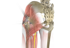
Hip Anatomy
The hip joint is the largest weight-bearing joint in the human body. It is also referred to as a ball and socket joint and is surrounded by muscles, ligaments and tendons. The thighbone or femur and the pelvis join to form the hip joint.
Any injury or disease of the hip will adversely affect the joint's range of motion and ability to bear weight.
The hip joint is made up of the following:
- Bones and joints
- Ligaments of the joint capsule
- Muscles and tendons
- Nerves and blood vessels that supply the bones and muscles of the hip
Bones and Joints of the Hip
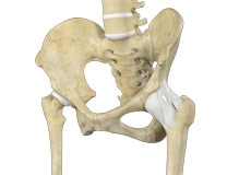
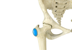
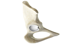
The hip joint is the junction where the hip joins the leg to the trunk of the body. It is comprised of two bones: the thighbone or femur, and the pelvis, which is made up of three bones called ilium, ischium and pubis.
The ball of the hip joint is made by the femoral head while the socket is formed by the acetabulum. The acetabulum is a deep, circular socket formed on the outer edge of the pelvis by the union of three bones: ilium, ischium and pubis. The lower part of the ilium is attached by the pubis while the ischium is considerably behind the pubis. The stability of the hip is provided by the joint capsule or acetabulum and the muscles and ligaments that surround and support the hip joint.
The head of the femur rotates and glides within the acetabulum. A fibrocartilaginous lining called the labrum is attached to the acetabulum and further increases the depth of the socket.
The femur is one of the longest bones in the human body. The upper part of the thighbone consists of the femoral head, femoral neck, and greater and lesser trochanters. The head of the femur joins the pelvis (acetabulum) to form the hip joint. Next to the femoral neck, there are two protrusions known as greater and lesser trochanters which serve as sites of muscle attachment.
Articular cartilage is the thin, tough, flexible and slippery surface lubricated by synovial fluid that covers the weight-bearing bones of the body. It enables smooth movements of the bones and reduces friction.
Ligaments of the Hip Joint
Ligaments are fibrous structures that connect bones to other bones. The hip joint is encircled with ligaments to provide stability to the hip by forming a dense and fibrous structure around the joint capsule. The ligaments adjoining the hip joint include:
- Iliofemoral ligament: This is a Y-shaped ligament that connects the pelvis to the femoral head at the front of the joint. It helps in limiting over-extension of the hip.
- Pubofemoral ligament: This is a triangular shaped ligament that extends between the upper portion of the pubis and the iliofemoral ligament. It attaches the pubis to the femoral head.
- Ischiofemoral ligament: This is a group of strong fibers that arise from the ischium behind the acetabulum and merge with the fibers of the joint capsule.
- Ligamentum teres: This is a small ligament that extends from the tip of the femoral head to the acetabulum. Although it has no role in hip movement, it does have a small artery within that supplies blood to a part of the femoral head.
- Acetabular labrum: The labrum is a fibrous cartilage ring which lines the acetabular socket. It deepens the cavity increasing the stability and strength of the hip joint.
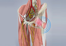
Muscles and Tendons of the Hip Joint
A long tendon called the iliotibial band runs along the femur from the hip to the knee and serves as an attachment site for several hip muscles including the following:
- Gluteal: These are the muscles that form the buttocks. There are three muscles (gluteus minimus, gluteus maximus, and gluteus medius) that attach to the back of the pelvis and insert into the greater trochanter of the femur.
- Adductors: These muscles are in the thighs which help in adduction, the action of pulling the leg back towards the midline.
- Iliopsoas: This muscle is in front of the hip joint and provides flexion. It is a deep muscle that originates from the lower back and pelvis, and extends up to the inside surface of the upper part of the femur.
- Rectus femoris: This is the largest band of muscles located in front of the thigh. They are also called hip flexors.
- Hamstring muscles: These begin at the bottom of the pelvis and run down the back of the thigh. Because they cross the back of the hip joint, they help in extension of the hip by pulling it backwards.
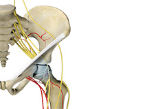
Nerves and Arteries of the Hip Joint
Nerves of the hip transfer signals from the brain to the muscles to aid in hip movement. They also carry the sensory signals such as touch, pain, and temperature back to the brain.
The main nerves in the hip region include the femoral nerve in the front of the femur and the sciatic nerve at the back. The hip is also supplied by a smaller nerve known as the obturator nerve.
In addition to these nerves, there are blood vessels that supply blood to the lower limbs. The femoral artery, one of the largest arteries in the body, arises deep in the pelvis and can be felt in front of the upper thigh.
Hip Movements
All the anatomical parts of the hip work together to enable various movements. Hip movements include flexion, extension, abduction, adduction, circumduction, and hip rotation.
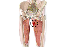
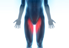
Hip Adductor Injuries
Hip adductors are the group of muscles on the inner side of your thigh that enable adduction or the ability to bring the thighs together.
Hip Ligament Injuries
Injuries to the hip ligaments are commonly called a hip sprain and can range from minor tears of the ligaments to more serious injuries involving the hip muscles, tendons or bone.
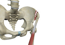
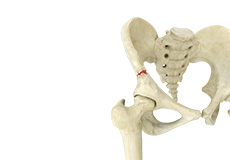
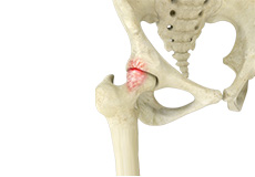
Avulsion Fractures of the Pelvis
An avulsion fracture of the pelvis is an injury that can occur when a tendon or ligament pulls off a piece of bone from the area surrounding the hip. This results in a part of the pelvic (hip) bone breaking away from the main part of the bone.
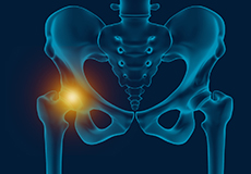
Hip Injury
The hip joint is one of the most important and flexible joints in the human body which allows us to walk, run, bend and perform physical activities.
Snapping Hip Syndrome
Snapping hip syndrome is a condition in which you hear or feel a snapping sound in the hip when you swing your legs, run, walk or get up from a chair. The sound can be experienced in the back, front or side of the hip.
Pelvic Fractures
A pelvic fracture is a condition that occurs due to the breakage of the pelvic bone. It may cause damage to the internal organs, nerves and blood vessels associated with the pelvic region.
Hip Bursitis
Hip bursitis is a painful condition caused by the inflammation of a bursa in the hip. Bursae are fluid-filled sacs present in the joints between bone and soft tissue to reduce friction and provide cushioning during movement.
Femoroacetabular Impingement
Femoroacetabular impingement (FAI) is a condition characterized by excessive friction in the hip joint from the presence of bony irregularities. These cause pain and decreased range of hip motion.
Avascular Necrosis
Avascular necrosis, also called osteonecrosis, is a condition in which bone death occurs because of inadequate blood supply to it. Lack of blood flow may occur when there is a fracture in the bone or a joint dislocation that may damage nearby blood vessels.
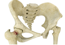
Hip Fracture
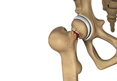
The hip joint is a “ball and socket” joint. The “ball” is the head of the femur or thighbone, and the “socket” is the cup-shaped acetabulum. The joint surface is covered by a smooth articular surface that allows pain-free movement in the joint.
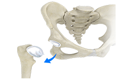
Hip Dislocation
The hip joint is a “ball and socket” joint. The “ball” is the head of the femur or thighbone, and the “socket” is the cup-shaped acetabulum. The joint is surrounded by muscles, ligaments, and tendons that support and hold the bones of the joint in place.
Hip Labral Tear
A hip labral tear is an injury to the labrum, the cartilage that surrounds the outside rim of your hip joint socket.
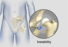
Hip Instability
Injury or damage to these structures can lead to a condition called hip instability when the joint becomes unstable.
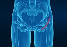
Hip Groin Disorders
Hip and groin disorders are more common in athletes. They are caused by rapid acceleration and deceleration motion.
Transient Osteoporosis of the Hip
Transient osteoporosis of the hip is a rare condition that causes temporary bone loss in the upper region of the thighbone (femur). It occurs most often in young or middle-aged men of the age groups 30 to 60, and women in their later stages of pregnancy or early postpartum (following childbirth).
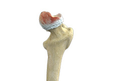
Osteoarthritis of the Hip
Osteoarthritis, also called degenerative joint disease, is the most common form of arthritis. It occurs most often in the elderly. This disease affects the tissue covering the ends of bones in a joint called cartilage.
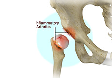
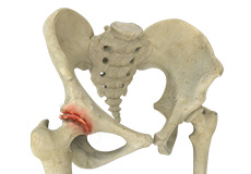
Inflammatory Arthritis of the Hip
The inflammation of the joints is referred to as arthritis. Inflammation arises when the smooth lining called cartilage at the ends of bones wears away.
Groin Injuries in Athletes
Groin injuries are injuries sustained by athletes during sports activity. Groin injuries comprise about 2 to 5 percent of all sports injuries. The most common kind of groin injury is a groin strain or a pulled groin muscle.
Hip Osteonecrosis
Hip osteonecrosis occurs due to disruption of the blood supply to the highest part of the thigh bone (femoral head). Due to lack of nourishment, the bone tissue of the femoral head dies and gradually collapses, which may further lead to degeneration of the underlying cartilage.
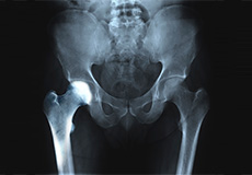
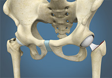
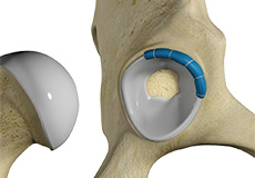
Outpatient Hip Replacement
Hip replacement surgery is one of the most common orthopedic surgeries performed. It involves the replacement of the damaged hip bone (ball shaped upper end of the femur) with a ceramic ball attached to a metal stem that is fixed into the femur and placing a new cup with a special liner in the pelvis.
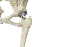
Total Hip Replacement
Total hip replacement is a surgical procedure in which the damaged cartilage and bone is removed from the hip joint and replaced with artificial components.
Hip Hemiarthroplasty
Hip hemiarthroplasty is a surgical technique employed to treat hip fractures. In this procedure, only one half (ball section) of the hip joint is substituted by a metal prosthesis.
Minimally Invasive Total Hip Replacement
Hip arthritis is a painful and common disease of the hip joint caused by damage to the cartilage. Total hip replacement surgery is an option to relieve severe arthritis pain that limits your daily activities.
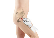
Anterior Hip Replacement
Anterior hip replacement surgery is performed under general anesthesia or regional anesthesia. You will lie down on your back, on a special operating table that enables your surgeon to perform the surgery from the front of the hip.
Outpatient Anterior Approach Hip Replacement
With improved technology and advances in anesthesia and pain control, hip replacement surgery has evolved and is now being offered in an outpatient setting.
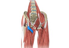
Posterior Hip Replacement
Posterior hip replacement is a minimally invasive hip surgery performed to replace the hip joint. It is also referred to as muscle sparing surgery because no muscles are cut to access the hip joint, enabling a quicker return to normal activity.
Hip Trauma Reconstruction
Hip trauma is an injury in the hip due to the impact caused by incidents such as a car accident or a hard fall. The injury can be a bone break or dislocation or both.